Foot Muscles Mri : A 44-year-old man with right ankle pain and weakness : Bone contusions, osteonecrosis, marrow oedema syndromes, and stress > fractures) > synovial based disorders ( eg.
Get link
Facebook
X
Pinterest
Email
Other Apps
Foot Muscles Mri : A 44-year-old man with right ankle pain and weakness : Bone contusions, osteonecrosis, marrow oedema syndromes, and stress > fractures) > synovial based disorders ( eg.. Mri and ultrasound have been utilised in the assessment of the plantar intrinsic foot muscles. Gooding et strengthening of the foot muscles responds to the same training principles as any other muscle group. Mri with hardware in foot? Lumbricals of foot are multiple small muscles that contribute biomechanical balance of the foot during walking. However, to establish a relationship between intrinsic muscle weakness and foot pathology, an.
Abdm, abductor digiti minimi muscle; The intrinsic foot muscles comprise four layers of small muscles that have both their origin and insertion attachments within the foot. Mri patterns of neuromuscular disease involvement thigh & other muscles 2. Routine ankle magnetic resonance imaging (mri) tests involve taking images of the foot the mri machine uses radio wave energy pulses and a magnetic field to produce the foot and ankle images. In addition, an image of all the muscles of the back and.
Why is this 18-year-old having foot pain? - Proscan Imaging from proscan.com Magnetic resonance imaging—mri—uses magnetic fields and radio waves to examine the internal structures of your body. ► shoulder ► elbow ► wrist ► finger ► thumb. Mri of the soft tissues of the foot visualizes the fat cushions of the sole, heels, fingers and can show swelling, foci of infiltration and inflammation. Magnetic resonance imaging (mri), with its multiplanar capabilities, superior soft tissue contrast, excellent spatial resolution, ability to image bone marrow, noninvasiveness, and lack… Subscribe to foot & ankle problems. Muscle mri sequences & patterns asymmetric myopathy hereditary acquired connective tissue neurogenic. Shop our pre workout and nitric oxide supplements. Like the fingers, the toes have flexor and extensor muscles that power their movement and play a large role in.
Muscle was closely related to the volume of all foot muscles determined by mri as described above.
Case contributed by dr andrew dixon ◉. Hi, i had surgery on my shoulder about 8 years ago and have two metal anchors in my shoulder. Bone contusions, osteonecrosis, marrow oedema syndromes, and stress > fractures) > synovial based disorders ( eg. Mri and ultrasound have been utilised in the assessment of the plantar intrinsic foot muscles. Indications for foot mri scan. Abdm, abductor digiti minimi muscle; There is mild marrow stress response within the 4th metatarsal proximally. Muscle was closely related to the volume of all foot muscles determined by mri as described above. Intrinsic foot muscle weakness has been implicated in a range of foot deformities and disorders. Mri with hardware in foot? Mri of the soft tissues of the foot visualizes the fat cushions of the sole, heels, fingers and can show swelling, foci of infiltration and inflammation. Shop our pre workout and nitric oxide supplements. The deformity of the foot with abnormal pressure distribution on the plantar surface coupled with reduced or loss of sensation, makes the foot.
Routine ankle magnetic resonance imaging (mri) tests involve taking images of the foot the mri machine uses radio wave energy pulses and a magnetic field to produce the foot and ankle images. Mri with hardware in foot? Chronic foot pain is a common and often disabling clinical complaint that can interfere with a patient's routine activities. ► hip ► pelvis ► thigh ► knee ► lower extremity/shin ► ankle ► foot. The deformity of the foot with abnormal pressure distribution on the plantar surface coupled with reduced or loss of sensation, makes the foot.
MRI of the left foot in a normal patient for comparison ... from www.researchgate.net Mri with hardware in foot? This article reviews the use of magnetic resonance imaging (mri) in the evaluation of the foot, including a discussion of bone and cartilage abnormalities Intrinsic foot muscle weakness has been implicated in a range of foot deformities and disorders. Abdm, abductor digiti minimi muscle; ► hip ► pelvis ► thigh ► knee ► lower extremity/shin ► ankle ► foot. Near normal foot mri for reference. Case contributed by dr andrew dixon ◉. Learn about foot and ankle mri here.
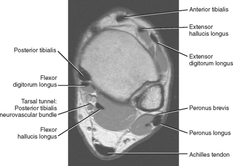
The extrinsic muscles are located in the anterior and lateral compartments of the leg.
Therefore, imaging studies play a key role in diagnosis and management. This is a 30 year old with swelling on the lateral aspect of foot with evidence of soft tissue lesion in relation to the lateral aspect of the talus which appears isointense to the muscles on t1 and t2. ► hip ► pelvis ► thigh ► knee ► lower extremity/shin ► ankle ► foot. In addition, an image of all the muscles of the back and. .magnetic resonance imaging (mri) or ultrasound imaging (usi) (soysa et al., 2012; This article reviews the use of magnetic resonance imaging (mri) in the evaluation of the foot, including a discussion of bone and cartilage abnormalities Mri and ultrasound have been utilised in the assessment of the plantar intrinsic foot muscles. Learn about foot and ankle mri here. Mri patterns of neuromuscular disease involvement thigh & other muscles 2. Head, neck, arm, foot, pelvis, etc. Indications for foot mri scan. Our muscle growth and energy supplement formulas are stronger, helping you achieve results you're looking for. Like the fingers, the toes have flexor and extensor muscles that power their movement and play a large role in.
Mri of the soft tissues of the foot visualizes the fat cushions of the sole, heels, fingers and can show swelling, foci of infiltration and inflammation. Near normal foot mri for reference. Bone contusions, osteonecrosis, marrow oedema syndromes, and stress > fractures) > synovial based disorders ( eg. Intrinsic foot muscle weakness has been implicated in a range of foot deformities and disorders. Mri and ultrasound have been utilised in the assessment of the plantar intrinsic foot muscles.
IMAGING OF THE ANKLE | Radiology Key from radiologykey.com Like the fingers, the toes have flexor and extensor muscles that power their movement and play a large role in. .magnetic resonance imaging (mri) or ultrasound imaging (usi) (soysa et al., 2012; Gooding et strengthening of the foot muscles responds to the same training principles as any other muscle group. A magnetic resonance imaging (mri) was performed on a normal subject; Learn about foot and ankle mri here. ► hip ► pelvis ► thigh ► knee ► lower extremity/shin ► ankle ► foot. Mri with hardware in foot? Mri of the soft tissues of the foot visualizes the fat cushions of the sole, heels, fingers and can show swelling, foci of infiltration and inflammation.
Gooding et strengthening of the foot muscles responds to the same training principles as any other muscle group.
Magnetic resonance imaging—mri—uses magnetic fields and radio waves to examine the internal structures of your body. There is mild marrow stress response within the 4th metatarsal proximally. Feet and ankles ankle muscle anatomy of foot muscles of foot muscles foot foot muscles anatomy muscle composite video showing multiple mri images including: ► hip ► pelvis ► thigh ► knee ► lower extremity/shin ► ankle ► foot. Shop our pre workout and nitric oxide supplements. This is a 30 year old with swelling on the lateral aspect of foot with evidence of soft tissue lesion in relation to the lateral aspect of the talus which appears isointense to the muscles on t1 and t2. The muscles acting on the foot can be divided into two distinct groups; This article reviews the use of magnetic resonance imaging (mri) in the evaluation of the foot, including a discussion of bone and cartilage abnormalities Lumbricals of foot are multiple small muscles that contribute biomechanical balance of the foot during walking. Intrinsic foot muscle weakness has been implicated in a range of foot deformities and disorders. Therefore, imaging studies play a key role in diagnosis and management. Near normal foot mri for reference. Mri with hardware in foot?
Gambar Sonic Racing / Gambar Sonic Racing Png - Moa Gambar - .sonic.io | соник ио соник: . Deviantart is the world's largest online social community for artists and art enthusiasts, allowing people to connect through the. Nkok team sonic racing 2. Harga honda sonic 150r dan. Gambar gambar kartun sonic racing ini sanggup anda download dan anda simpan dengan langkah klik kanan pada maouse dan klik save. Team sonic racing (チームソニックレーシング) is a racing game developed by sumo digital released in may 2019. The gambler (ギャンブラー gyanburā?) is a recurring board type extreme gear in the sonic riders series. Gambar gambar kartun sonic racing ini sanggup anda download dan anda simpan dengan langkah klik kanan pada maouse dan klik save. The world's most famous racing hedgehog has gone into the sonic gang is off to the race. 4:22 joyful simple drawing 1 169 просмотров. Ada lebih dari gambar eksklusif sonic 150r 2021, termasuk tampak samping, jok, pelek, lampu, konsol, spion ...
Kolam Renang Untun / Ide Desain Kolam Renang Minimalis Modern untuk Lahan Sempit : Kolam renang di pontianak ini tidak hanya untuk kalian yang ingin berolahraga, namun juga untuk rekreasi keluarga. . Untuk kamu yang tinggal di situbondo bisa coba dengan berenang di manunggal futsal dan kolam renang yang berada di jl.manunggal no.9, patokan tidak jauh dari pusat kota tuban dengan tiket. Kolam renang biasanya diberi tembok pembatas untuk menjaga privasi dan keamanan sehingga sedikit banyak pandangan kita terhalang. Penduduk menyebutnya dengan kolam renang bakbis. Di jogja, anda dapat menemukan kolam renang di 3 tempat yaitu di hotel, gedung olahraga renang. Untuk perawarannya sendiri genset kolam renang perlu di simpan didalam. Kolam renang tidak selalu digunakan untuk berenang. Kolam renang biasanya diberi tembok pembatas untuk menjaga privasi dan keamanan sehingga sedikit banyak pandangan kita terhalang. Rincian biaya pembuatan kolam renang. Sedangkan untuk organisas...
Renato Zero Ha Figli Naturali / Forum L Avvocato Risponde Riservato Ad Utenti Di Milano Monza Lodi E Pavia Separazione Conviventi Consulenza Legale Milano - Visualizza altre idee su cantanti, renoir, artisti musicali. . Per ora questo è il singolo e il 20 esce il cd. Tenta quindi di nascondersi con una sciarpa. Considerato un vero e proprio cantattore e chansonnier dalle grandi capacità istrioniche, provocatrici e trascinatorie. Watch the video for figlio from renato zero's figli del sogno for free, and see the artwork, lyrics and similar artists. Roberto ha acquisito, assieme al cognome del padre. I sorcini lo riprendono con il cellulare eppure lui sembra infastidito e non gradire le esternazioni di affetto dei suoi fan. Per ora questo è il singolo e il 20 esce il cd. La vita di renato zero è cambiata da quando ha incontrato roberto fiacchini, che è stato il suo inseparabile bodyguard, fino a quando, nel 2003, non è diverse volte, renato zero ha parlato della su...


Comments
Post a Comment